


Tips On Finding The Best Business Coach
Finding the perfect business coach is essential for entrepreneurs and professionals looking to grow their
Finding the perfect business coach is essential for entrepreneurs and professionals looking to grow their

Dental crowns have become increasingly popular due to their versatility and effectiveness in addressing various

Achieving a cohesive and harmonious interior design involves combining various elements to create a unified

Embarking on the journey of renovating your dream home is an exciting and transformative endeavor.

Homework is a vital part of the learning process, providing students with an opportunity to
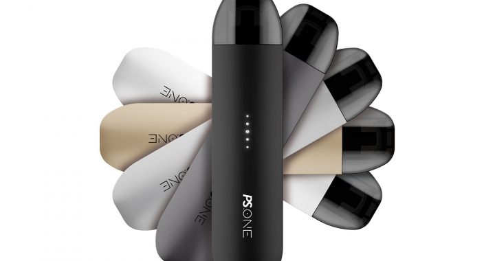
Vaping has become a prevalent practice, and like any social activity, it comes with its

A gent’s salon is not just a place for a haircut or grooming; it’s an
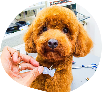
Your dog’s coat is a prominent part of its appearance, and a well-groomed coat not

Hydraulics is a technology that utilizes the power of fluid pressure to perform mechanical tasks.

In the world of welding, gas regulators play a vital role in ensuring the smooth